Lab 9: Organ Systems (fetal pig dissection)
Preparation for lab
To get the most out of this lab you need to be prepared. The basic knowledge needed for this lab is covered in Amerman “Human Anatomy and Physiology” in Chapter 1.2 “Overview of Anatomy and Physiology”.
Introduction
Now that we have worked our way through several organ systems in the body, it is time to revisit the internal anatomy through dissection. Since we do not have human cadavers, we will be using an anatomically closely related species to allow you to explore the thoracic, abdominal, and pelvic cavities as they pertain to the cardiovascular, respiratory, digestive and, to a lesser extent, integumentary and lymphatic systems. The internal anatomy of a pig is very similar in construction to a that of humans. Please remember that your pig was once a living organism and should be treated with respect and care.
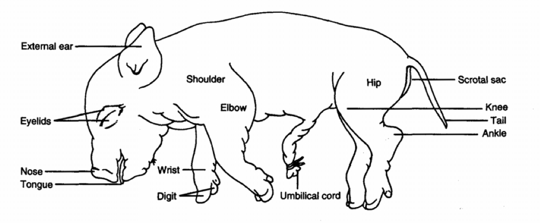
External anatomy of a fetal pig.
- Examine the fetal pig and locate the external features shown in the image.
- Locate the two rows of nipples of mammary glands which are present on the ventral abdominal surface of both males and females. Mammary glands later develop only in maturing females.
- As we are looking at a fetal pig, there is an umbilical cord: Make a transverse cut through the umbilical cord and examine the cut end. Locate the two umbilical arteries that carry blood from the fetal pig to the placenta, and the single umbilical vein that delivers nutrient-rich blood back to the fetal pig.
- Determine the sex of your specimen: The female pig has an urogenital opening immediately ventral to the anus and has a small genital papilla marking its location. The male pig shows a scrotal sac ventral to the anus and the urogenital opening is just posterior to the umbilical cord.
External Anatomy: Upper Respiratory System
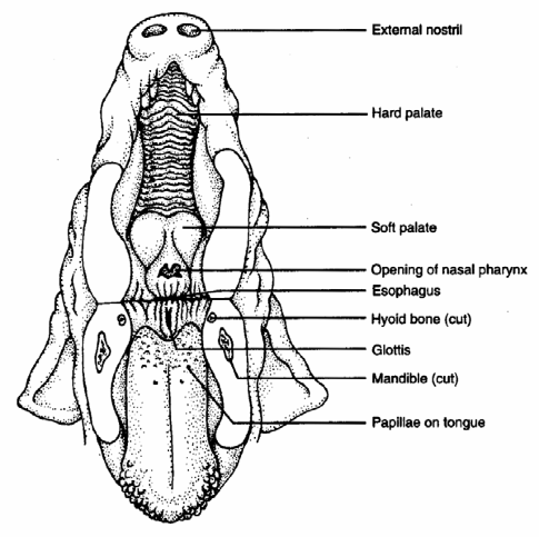
To expose the structures of the mouth and pharynx, start by inserting a pair of scissors in the corner of the mouth and cut posteriorly through the cheek. Open the mouth as you make your cut and follow the curvature of the tongue to avoid cutting the roof of the mouth.
Hold down the epiglottis and surrounding tissue and continue your incision dorsal to it and on into the opening of the esophagus. Now, repeat the procedure on the other side so that the lower jaw can be pulled down to identify the following structures:
To see the remaining systems you need to dissect the pig. This diagram shows the correct order and location of incisions.
- Teeth (deciduous teeth may have erupted)
- Tongue - Papillae
- Hard and soft palate
- Nasopharynx
- Oropharynx
- Laryngopharynx
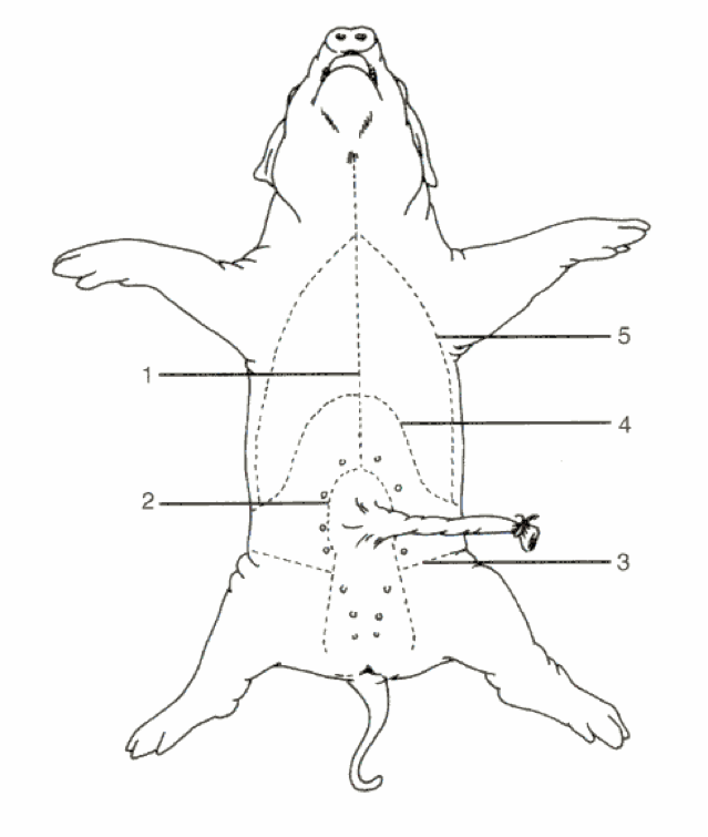
In order to see the remaining systems, you will need to dissect your pig. PLEASE BE CAREFUL NOT TO INJURE YOURSELF! Make incisions along the lines shown in the figure above, but keep the cutting to a minimum: “to dissect” does not mean “to cut up”, but rather “to expose to view”! If done right, your dissection should look like the one shown in the picture below. Use plenty of pins to spread the preparation open, then begin to identify the individual organ systems.
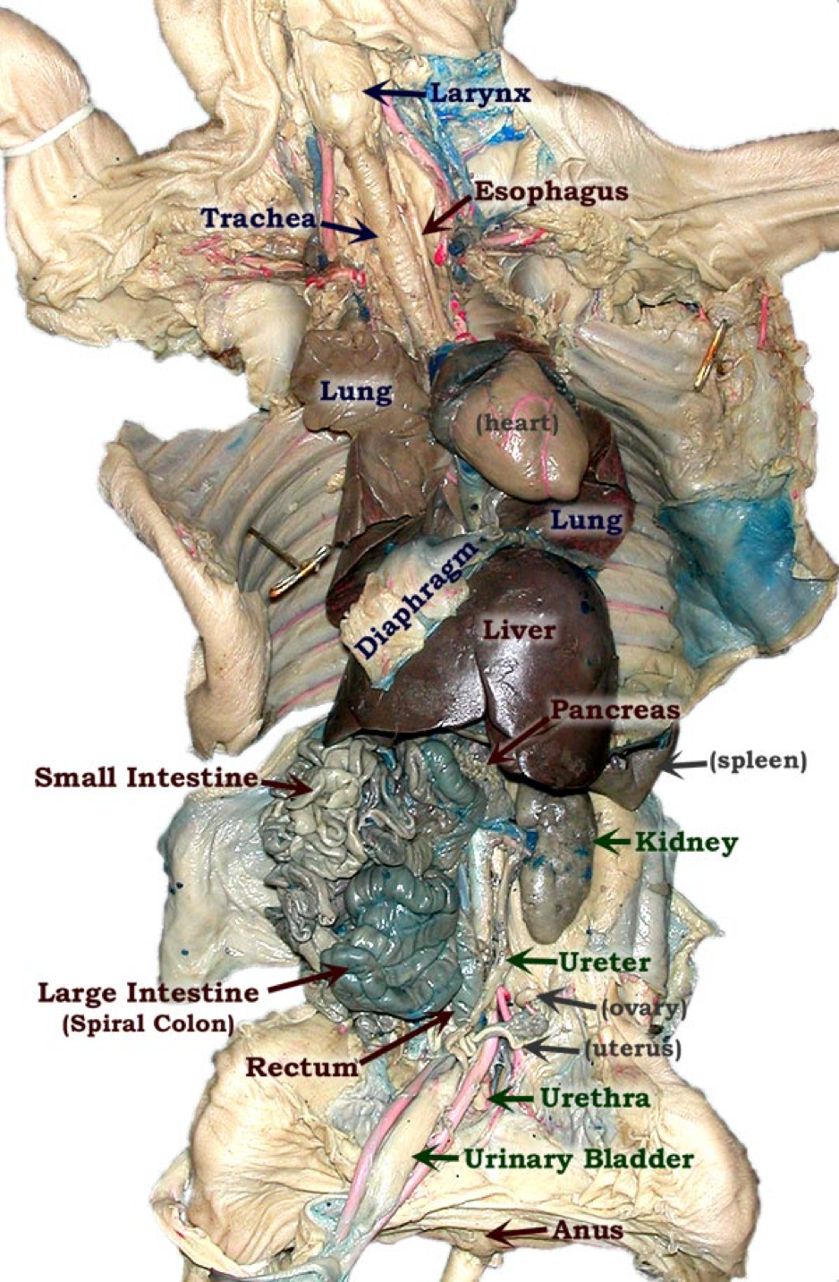
Open thoracic, abdominal, and pelvic cavity, with major organ systems labeled.
Thoracic Cavity
The thoracic cavity is protected by the rib cage and contains the pleural cavity containing the lungs as well as the mediastinum containing the heart. Note the 4 lobes of the lungs and how they hang relatively loose in the pleural cavity, mostly attached by the primary bronchi. As we are dissecting fetal pigs, the lungs were never inflated and are thus not fully developed.
Locate and study:
- Trachea with cartilaginous rings (why does it have these rings?)
- Lungs (four lobes)
- Diaphragm
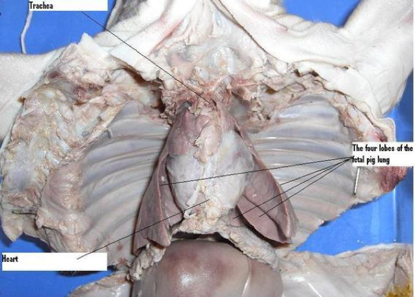
As we are dissecting fetal pigs, the lungs were never inflated or functional. Many students are surprised by how small they are.
Mediastinum of the thoracic cavity: the pericardial cavity
The pericardial cavity is surrounded by the pulmonary cavity to either side and contains the heart. Note how the heart is only attached at its base and can slide around (in order to properly see the heart you may need to remove parts of the thymus gland). Try to follow the blood vessels as well as you can. Open up the heart and try to compare it to the adult pig heart you previously dissected.
Locate and study:
- pericardial sac
- inferior and superior vena cava
- pulmonary trunk and veins
- coronary blood vessels
- heart chambers

The heart with clearly visible coronary blood vessels.

Abdominopelvic Cavity
The abdominal cavity contains most of the digestive, the urinary, and the reproductive systems. The stomach, small intestine, pancreas, and spleen are held in place by the mesentary - double-folds of the peritoneum and since 2017 considered an organ! It provides mechanical stability, but also blood and lymph vessels, nerves, and storage for fat.
Locate and study:
- Stomach
- Liver and hepatic portal vein
- Spleen
- Gall bladder and bile duct
- Pancreas
- Small intestine: Duodenum, Jejunum, Illeum
- Appendix
- Large intestine: Haustral pouches, Ascending colon, Transverse colon, Descending colon
- Kidney with renal artery and vein, and ureter
- Urinary bladder
- Reproductive organs: ovaries, uterus, vagina in female pig, testes (undescended in younger, in scrotum in older fetuses), prostate & bulbourethal gland in male pigs
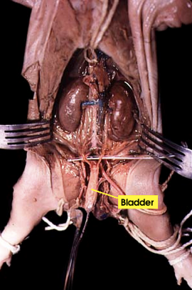
The abdominal cavity is often grouped with the pelvic cavity and referred as abdominopelvic cavity. Shown here is the lower part with the bladder labeled.
Setup & supplies
For each table
- fetal pig
- pins
- dissection dish
- dissection tools (scalpel, forceps, scissors, probe)
- gloves
- wash bottle
For the lab
- black plastic bags for disposal of hearts
- spray bottle with EtOH to wipe down tables