Lab 11: Kidney and Urine Analysis
Preparation for lab
To get the most out of this lab you need to be prepared. The basic knowledge needed for this lab is covered in Chapter 24 “The Urinary System”
Introduction
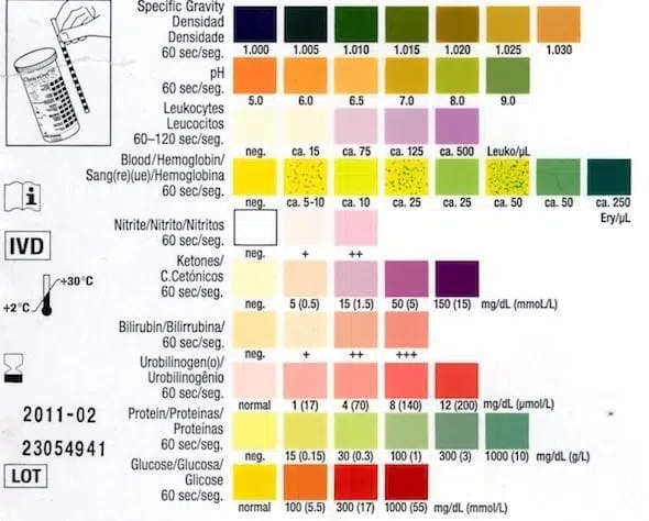
A urinalysis (UA) is often performed by your physician as part of a routine medical exam to screen for early signs of disease. If you have signs of diabetes or kidney disease, to monitor you if you are being treated for these conditions, to check for blood in the urine, or to diagnose a urinary tract infection. A part of a urinalysis can be performed by using urine test strips, in which the test results can be read as color changes.
Physiology
DIRECTIONS:
-
go to bathroom and urinate into a cup
-
make observations on physical characteristics
-
dip test strip into urine, blot excess with paper towel, and wait appropriate amount of time for results (see packaging)
-
record results on the table and compare to normal values
| test | expected result | your result |
|---|---|---|
| volume | 40 ml / hr | |
| transparency | transparent | |
| pH | 4.5 - 8 | |
| specific gravity | 1.001 - 1.030 | |
| leucocytes | negative | |
| nitrite | negative | |
| urobilinogen | negative | |
| protein | negative | |
| blood | negative | |
| ketone | negative | |
| bilirubin | negative | |
| glucose | negative |
Test strips provide a quick and cheap method to screen for medical conditions, they are, however, not suitable to diagnose (i.e. they are not reliable). A positive result on the test strip might indicate might indicate further diagnostic tests.
- pH: The acidity/alkalinity of urine. Abnormal pH levels can be associated with kidney stones, UTIs, or kidney disorders.
-
Specific Gravity: Measures urine concentration. Abnormal levels can indicate conditions such as dehydration, kidney disorders, or inappropriate ADH (antidiuretic hormone) secretion.
- Leukocytes: The presence of white blood cells in urine might indicate a urinary tract infection (UTI) or kidney infection.
- Nitrites: Bacteria that cause UTIs can convert nitrates into nitrites. A positive nitrite test can be an indicator of a UTI.
- Urobilinogen: Elevated levels can indicate liver diseases such as hepatitis or cirrhosis, or conditions causing hemolysis (breakdown of red blood cells).
- Protein: Protein in urine (proteinuria) can be a sign of kidney damage or disease, high blood pressure, or other medical conditions.
- Blood: The presence of blood can indicate kidney stones, UTIs, kidney injury, or bladder cancer.
- Ketones: The presence of ketones can indicate diabetic ketoacidosis (a complication of diabetes), starvation, or a carbohydrate-deficient diet.
- Bilirubin: The presence of bilirubin can suggest liver disease such as hepatitis, cirrhosis, or bile duct obstruction.
- Glucose: Glucose in urine (glucosuria) can be a sign of diabetes mellitus or a condition leading to excessive blood sugar levels.
Anatomy
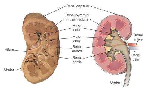
What can you deduct from the anatomy of a kidney about how it works? What are the kidneys and what do they do? The kidneys are two bean-shaped organs, each about the size of a fist. They are located just below the rib cage, one on each side of the spine. Every day, the two kidneys filter about 120 to 150 quarts of blood to produce about 1 to 2 quarts of urine, composed of wastes and extra fluid. The urine flows from the kidneys to the bladder through two thin tubes of muscle called ureters, one on each side of the bladder. The bladder stores urine. The muscles of the bladder wall remain relaxed while the bladder fills with urine. As the bladder fills to capacity, signals sent to the brain tell a person to find a toilet soon. When the bladder empties, urine flows out of the body through a tube called the urethra, located at the bottom of the bladder. In men the urethra is long, while in women it is short. Assume you are the first scientist to try to figure out how the kidney does what it does: Filter blood and excrete the waste products as urine.
DIRECTIONS:
- Carefully dissect the kidney in front of you, paying close attention to texture and color differences.
- Make a sketch of the kidney that will help you to explain how the kidney works.
Food for thought:
-
Where does “dirty” blood enter the kidney?
-
Where does “clean” blood leave the kidney?
-
What is urine and where does it leave the kidney?
-
What happens in the renal cortex?
-
The medulla is the region located inward from the cortex. What happens here?
Setup & supplies
On each table
-
dissection dish with kidney
-
dissection tools
On side bench
-
gloves
-
googles
-
plastic cups
-
URS10 test strips